IMRT Pelvic 3D Phantom
COMPLETE QA FROM CT IMAGING TO DOSE VERIFICATION
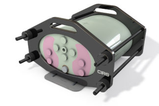
The CIRS Model 002PRA IMRT Pelvic 3D Phantom for film and ion chamber dosimetry is designed to address the complex issues surrounding commissioning and comparison of treatment planning systems and complete system QA from CT imaging to dose verification.
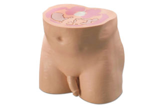
Our CIRS Pelvic 3D Phantom properly represents human pelvic anatomy in shape, proportion and structure as well as density. This enables thorough analysis of both the imaging and dosimetry system. Elliptical in shape, the phantom approximates the size of an average patient and has a tissue equivalent three-dimensional skeleton. It’s constructed of proprietary tissue equivalent epoxy materials.
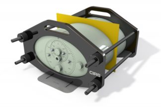
The Model 002PRA includes four different Electron Density reference plugs which can be interchanged in five separate locations within the phantom. The surface of the phantom is etched with grooves to ensure proper orientation of the CT slices and accurate film to plan registration.
Features:
- Verify heterogeneity corrections
- Correlate CTU to electron density
- Check dose distributions in sensitive areas
- Check depth doses and absolute dose
- 2D and 3D isodoses
- Calibrate film with ion chamber*
- Verify individual patient treatment plans
The CIRS line of IMRT phantoms is compatible with the RIT 113 software for film to plan analysis. GafChromic ® is a registered trademark of International Specialty Products, Wayne, NJ.
NOTE: This product or an optional accessory of this product requires a CIRS dosimetry cavity code before an order can be placed. Please refer to the Dosimetry Cavity Codes document to identify the CIRS code for the probe you intend to use with this product.
| Aland, Trent J; 'Quality assurance of complex radiotherapy treatments using high-resolution 2D dosimeters'. 2022; Queensland University of Technology. View |
| Aland, Trent; Williams, Talia; Spalding, Myles; Kairn, Tanya; Trapp, Jamie; '7. Use of in vivo transit portal images to detect inter-fraction patient geometry changes on an O-ring type linear accelerator for pelvis and head/neck patients.'. Quality Assurance of Complex Radiotherapy Treatments using High-Resolution 2D Dosimeters. 2021; 149. Queensland University of Technology. View |
| Aland, Trent; Jones, Mark; Aho, Jari; Kairn, Tanya; Trapp, Jamie; '6. Modelling and theoretical improvements to the visualisation of implanted fiducial markers for intra-fraction MV imaging of prostate VMAT targets.'. Quality Assurance of Complex Radiotherapy Treatments using High-Resolution 2D Dosimeters. 2021; 122. Queensland University of Technology. View |
| Fleckenstein J, Jahnke L, Lohr F, Wenz F, Hesser J. Development of a Geant4 based Monte Carlo Algorithm to evaluate the MONACO VMAT treatment accuracy. Z Med Phys. 2013;23(1):33-45. View |
| Boggula R, Lorenz F, Abo-madyan Y, et al. A new strategy for online adaptive prostate radiotherapy based on cone-beam CT. Z Med Phys. 2009;19(4):264-76. View |
| Khosravi H, Hashemi B, Mahdavi SR, Hejazi P. Effect of Gold Nanoparticles on Prostate Dose Distribution under Ir-192 Internal and 18 MV External Radiotherapy Procedures Using Gel Dosimetry and Monte Carlo Method. J Biomed Phys Eng. 2015;5(1):3-14. View |
References
Please note: CIRS is now part of Sun Nuclear, making us stronger together. The full suite of CIRS solutions will remain available for browsing on this site and are also available for order through your Sun Nuclear sales representative.