Proton Therapy Dosimetry Head

Model 731-HN
TISSUE EQUIVALENT FOR PROTON AND PHOTON THERAPY

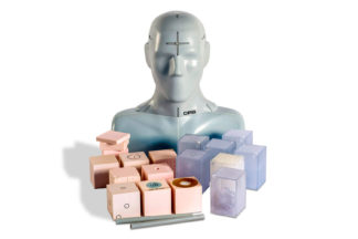
The CIRS Proton Therapy Dosimetry Head is an anthropomorphic head phantom designed for commissioning and treatment planning system (TPS) verification with any conformal or IMRT Proton Therapy system1.

The phantom is constructed of CIRS tissue-equivalent materials, which mimic reference tissues within 1.5% for protons and within 1% for photons from 50 KeV to 15 MeV. The Proton Therapy Head can be used during all standard IMRT procedures from CT image acquisition to proton beam delivery verification1,3.

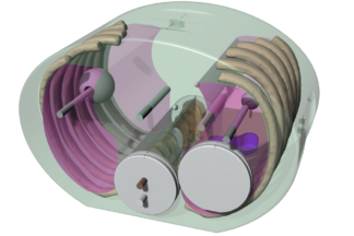
Tissue equivalency of detailed internal structures makes the phantom ideal for treatment plan evaluation in high density-gradient locations, which are specifically important in proton therapy2, such as air cavity vs. bone structures. Internal structures include brain, bone with cortical and trabecular distinction, larynx, trachea, fully-open sinus cavities, nasal and mouth cavities, and teeth with distinct dentine, enamel and root structure.

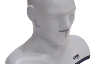
Model 731-HN approximates the average male human head in both size and structure to allow for intuitive set up with any patient positioning or fixation device.

One half of the phantom is sectioned in 2 cm increments for three film locations in the cranio-caudal direction starting from the approximate center of the sagittal plane. Because slices are orthogonal to the CT axial plane, artifacts caused by residual air gaps are noticeably reduced compared to standard axial slices.

Proton system commissioning is enhanced by placement of a tungsten BB in a molar and a titanium prosthesis attached by two screws at C3 and C5 vertebra.

Features:
  • Detailed internal anatomy including bone and air
  • Three film locations in sagittal direction
  • Tissue Equivalent for protons and photons
  • Dental filling and spine prothesis

Data Sheet

Proton Therapy Dosimetry Head: Data Sheet

Mein, Stewart; Kopp, Benedikt; Vela, Anthony; Dutheil, Pauline; Lesueur, Paul; Stefan, Dinu; Debus, Jürgen; Haberer, Thomas; Abdollahi, Amir; Mairani, Andrea; 'How can we consider variable RBE and LETd prediction during clinical practice? A pediatric case report at the Normandy Proton Therapy Centre using an independent dose engine'. Radiation Oncology. 2022; 17 (1): 16-Jan. BioMed Central. View
Gajewski, J; Garbacz, M; Chang, CW; Czerska, K; Durante, M; Krah, N; Krzempek, K; 'Commissioning of GPU-Accelerated Monte Carlo Code Fred for Clinical Applications in Proton Therapy'. Front. Phys. 8: 567300. doi: 10.3389/fphy. 2021; View
Rusanov, Branimir; Ebert, Martin Andrew; Mukwada, Godfrey; Hassan, Ghulam Mubashar; Sabet, Mahsheed; 'A convolutional neural network for estimating cone-beam CT intensity deviations from virtual CT projections'. Physics in Medicine & Biology. 2021; IOP Publishing. View
Nenoff, Lena; Matter, Michael; Charmillot, Marjolaine; Krier, Serge; Uher, Klara; Weber, Damien Charles; Lomax, Antony John; Albertini, Francesca; 'Experimental validation of daily adaptive proton therapy'. Phys Med Biol. 2021; View

Summary: A comprehensive and clinically applicable workflow for daily adaptive proton therapy is described that employs a custom head and neck phantom patterned off the 731-HN and the 038.
Civinini, Carlo; Scaringella, Monica; Brianzi, Mirko; Intravaia, Matteo; Randazzo, Nunzio; Sipala, Valeria; Rovituso, Marta; Tommasino, Francesco; Schwarz, Marco; Bruzzi, Mara; 'Relative stopping power measurements and prosthesis artifacts reduction in proton CT'. Physics in Medicine & Biology. 2020; 65 (22): 225012. IOP Publishing. View
Faller, F; Mein, S; Ackermann, B; Stiller, W; Mairani, A; 'Dosimetric validation of spectral CT-based stopping power prediction for proton beams using an anthropomorphic head phantom'. 2020; Heidelberg University Hospital. View
Salas Ramirez, Maikol; 'Methods to Improve Bone Marrow Dosimetry in Molecular Radiotherapy'. Julius-Maximilians-Universität Würzburg. View
Albertini F, Casiraghi M, Lorentini S, Rombi B, Lomax AJ. Experimental verification of IMPT treatment plans in an anthropomorphic phantom in the presence of delivery uncertainties. Phys Med Biol. 2011;56(14):4415-31.
Lorentini S, Menegotti L, Delana A, Schwarz M. PO-0873 DOSIMETRIC VERIFICATION OF IMRT PLANS WITH A CUSTOMIZED ANTHROPOMORPHIC PHANTOM. Radiotherapy and Oncology. 2012;103:S342.
Demez N, Lee T. EP-1320 STUDY ON THE COMPATIBILITY OF TISSUE EQUIVALENT PHANTOMS FOR USE IN PROTON BEAM THERAPY QA. Radiotherapy and Oncology. 2012;103:S501.
Huang JY, Kerns JR, Nute JL, et al. An evaluation of three commercially available metal artifact reduction methods for CT imaging. Phys Med Biol. 2015;60(3):1047-67.  View
Kaser, S; Bergauer, T; Birkfellner, W; Georg, D; Hatamikia, S; Hirtl, A; Irmler, C; Pitters, F; Ulrich-Pur, F; 'Image reconstruction for ion imaging with the TIGRE software framework'. View

Summary: This paper presents, TIGRE, a fast reconstruction method for proton CT, which was tested with the help of the 731-HN.

References

Model: 731-HN Modality: