Virtual Human Male Pelvis Phantom

Model 801-P
THE NEXT GENERATION IN ANTHROPOMORPHIC PHANTOMS

The CIRS Model 801-P phantom is our most realistic, tissue equivalent phantom available. The phantom was designed for use in diagnostic radiology and radiation therapy for imaging dosimetry teaching and demonstration applications. All anatomical dimensions of the phantom are based on The Visible Human Project (VHP) data sets that serve as a reference for the study of human anatomy and are available through the National Library of Medicine.

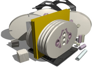
CIRS has combined this anatomical detail with our advanced tissue mimicking technology. The phantom is made from proprietary epoxy materials that mimic the density and radiation attenuation properties of human tissue within 1% from 50 keV to 25 MeV. It contains anatomically precise bone, cartilage, spinal cord, vertebral disks, muscle, intestines, bladder, prostate, rectum and interstitial fat.

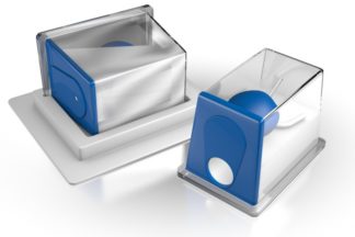
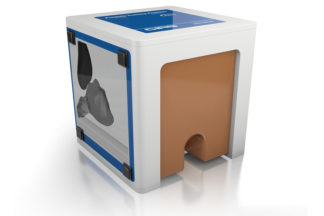
Just like the CIRS ATOM® line of phantoms, the VHP Phantom can be purchased uncut or segmented into 25mm thick contiguous sections. CIRS offers a hole grid options of 3 cm x 3 cm.

The grid contains 5mm diameter through holes filled with homogeneous tissue equivalent plugs for TLD placement. Hole locations are optimized for internal organ dosimetry. Ion chamber cavities, other grid patterns and hole diameters are available upon request.

Features:
  • Based on the Visible Human Project data set
  • Superior tissue simulation and lifelike imaging properties
  • Accommodates wide variety of detectors

Data Sheet

Virtual Human Male Pelvis Phantom: Data Sheet

Lehmann, Joerg; Standen, Therese S; Kaur, Guneet; Wolf, Joshua; Wilfert, Alex; Simpson, John; 'Methodology of thermal drift measurements for surface guided radiation therapy systems and clinical impact assessment illustrated on the C-Rad Catalyst+ HD system'. Technical Innovations & Patient Support in Radiation Oncology. 2022; 21: 58-63. Elsevier. View
Liu, L., Y. Han, and M. Jin. "Fast Alternating Projection Methods for Constrained Tomographic Reconstruction." Medical Imaging 2017: Ultrasonic Imaging and Tomography, 2017. Web.  View
Sykes, J., et al., Evaluation Report X-Ray Tomographic Image Guided Radiotherapy Systems. Radiotherapy Physics Group Medical Physics and Engineering & Radiotherapy Department, Non-Surgical Oncology. NHS Centre for Evidence-based Purchasing (UK).CEP 10071: March 2010. 
Sykes, J., et al., Protocol: Technical evaluation of X-ray tomographic image-guided radiotherapy devices. Radiotherapy Physics Group Medical Physics and Engineering & Radiotherapy Department, Non-Surgical Oncology. NHS Centre for Evidence-based Purchasing (UK). CEP10071: March 2010. 
Schaly B, Varchena V, Au P, Pang G. Evaluation of an anthropomorphic male pelvic phantom for image-guided radiotherapy. Reports in Medical Imaging. 2009; 2:69-78.  View
Schaly B, Varchena V, Au P, Pang G. Sci-Thur PM Therapy-05: Evaluation of male pelvic phantom for megavoltage cone-beam computed tomography. Medical Physics. 2006; 33(7).  View
Ouyang L, Song K, Solberg T, Wang J. A moving blocker system for cone-beam computed tomography scatter correction. Proc. SPIE 8668, Medical Imaging 2013: Physics of Medical Imaging, 86681P (March 6, 2013).  View
Ouyang L, Lee HP, Wang J. A moving blocker-based strategy for simultaneous megavoltage and kilovoltage scatter correction in cone-beam computed tomography image acquired during volumetric modulated arc therapy. Radiother Oncol. 2015;115(3):425-30.  View
Li H, Dolly S, Chen HC, et al. A comparative study based on image quality and clinical task performance for CT reconstruction algorithms in radiotherapy. J Appl Clin Med Phys. 2016;17(4):5763. 

References

Model: 801-P Modalities: ,