Smith, Blake R; Strand, Sarah A; Dunkerley, David; Flynn, Ryan T; Besemer, Abigail E; Kos, Jennifer D; Caster, Joseph M; Wagner, Bonnie S; Kim, Yusung; 'Implementation of a real‐time, ultrasound‐guided prostate HDR brachytherapy program'. Journal of Applied Clinical Medical Physics. 2021; View
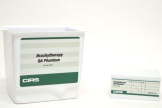
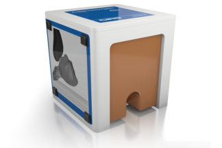
Summary: A workflow for commissioning of the Ocentra Prostate treatment planning system used in real-time, ultrasound-guided HDR brachytherapy is decribed. Image quality tests, as recommended in the AAPM Task Group 128 report, was performed with the Model 45B Brachytherapy QA phantom while end-to-end testing was performed with the Model 53L prostate phantom. |
| Doyle, Andrea J; Sullivan, Frank; Walsh, John; King, Deirdre M; Cody, Dervil; Browne, Jacinta E; 'Development and Preliminary Evaluation of an Anthropomorphic Trans-rectal Ultrasound Prostate Brachytherapy Training Phantom'. Ultrasound in Medicine & Biology. 2021; 47 (3): 833-846. Elsevier. View |
| Maris, Bogdan; Tenga, Chiara; Vicario, Rudy; Palladino, Luigi; Murr, Noe; De Piccoli, Michela; Calanca, Andrea; Puliatti, Stefano; Micali, Salvatore; Tafuri, Alessandro; 'Toward autonomous robotic prostate biopsy: a pilot study'. International Journal of Computer Assisted Radiology and Surgery. 2021; 9-Jan. Springer. View |
| Mahcicek, Davut Ibrahim; Yildirim, Korel D; Kasaci, Gokce; Kocaturk, Ozgur; 'Preliminary Evaluation of Hydraulic Needle Delivery System for Magnetic Resonance Imaging-Guided Prostate Biopsy Procedures'. Journal of Medical Devices. 2021; 15 (4): 41002. American Society of Mechanical Engineers. View |
| Mathur, Prateek; 'Transperineal ultrasound image guidance system for robot-assisted laparoscopic radical prostatectomy'. 2020; University of British Columbia. View |
| Morris, D Cody; Chan, Derek Y; Lye, Theresa H; Chen, Hong; Palmeri, Mark L; Polascik, Thomas J; Foo, Wen-Chi; Huang, Jiaoti; Mamou, Jonathan; Nightingale, Kathryn R; 'Multiparametric Ultrasound for Targeting Prostate Cancer: Combining ARFI, SWEI, QUS and B-Mode'. Ultrasound in Medicine & Biology. 2020; 46 (12): 3426-3439. Elsevier. View |
| Kemper J, Burkholder A, Jain A, et al. TU-EE-A1-06: Transrectal Fiducial Carrier for Radiographic Image Registration in Prostate Brachytherapy. Medical Physics. 2005; 32(6). View |
| Tremblay C, Gingras L, Archambault L, et al. SU-FF-T-232: Characterization and Use of MOSFET as In Vivo Dosimeters under 192Ir Irradiation for Real-Time Quality Assurance. Medical Physics. 2005; 32(6). View |
| Onik G, Downey D, Fenster A. Three-dimensional sonographically monitored cryosurgery in a prostate phantom. Journal of Ultrasound in Medicine. 1996; 15(3):267-270. View |
| Seifabadi, Reza. "TELEOPERATED MRIâ€GUIDED PROSTATE NEEDLE PLACEMENT." Thesis. Queen's University, Canada, 2013. View |
| Ukimura O, Desai MM, Palmer S, et al. 3-Dimensional elastic registration system of prostate biopsy location by real-time 3-dimensional transrectal ultrasound guidance with magnetic resonance/transrectal ultrasound image fusion. J Urol. 2012;187(3):1080-6. View |
| Wang, Y., D. Ni, M. Xu, X. Xie, and PA Heng. "Patient-specific Deformation Modelling via Elastography: Application to Image-guided Prostate Interventions." Scientific Reports, 07 June 2016. Web. View |
| Kim, ST, Y. Kim, and J. Kim. "Design of an MR-compatible Biopsy Needle Manipulator Using Pull-pull Cable Transmission." Korean Society for Precision Engineering, 10 Sept. 2016. Web. View |
| Fedorov, A., K. Tuncali, L. Panych, et al. "Segmented Diffusion-weighted Imaging of the Prostate: Application to Transperineal In-bore 3 T MR Image-guided Targeted Biopsy." Elsevier, 2016. Web. |
| Boroomand, A., E. Li, MJ Shafiee, et al . "A Unified Bayesian-based Compensated Magnetic Resonance Imaging." ResearchGate, 2016. Web. |
| Palladino, Luigi; Maris, Bogdan; Antonelli, Alessandro; Fiorini, Paolo; 'PROST-Net: A deep learning approach to support real-time fusion in prostate biopsy'. View |


